release time:2022-09-22 11:41:04
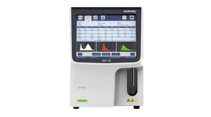
Many hospitals and clinics currently use 3-part and 5-part Differential Hematology Analyzers more often. The following is an introduction to the 3-part Differential Hematology Analyzers.

The 3-part Differential Hematology Analyzers use the instantaneous change in electrical resistance of blood cells passing through the micropores to generate a pulse current for counting. Dilute specimens of white and red blood cells are passed through their respective counting microwells and into their respective channels under the control of a negative pressure. The cell signals are then screened, amplified and counted by two photoelectric sensors.
a. Microtubes: A standard size ruby hole is set at its lower end. rbc tube hole; 80 microns; wbc tube hole diameter: 100 microns. Easy to clog, prevent plugging and maintenance.
b. Micron Counting: impurities other than cells will be mistaken for cell counting. Ensure the quality of the diluent and the cleanliness of the environment.
c. Cells can overlap each other through the micropores and produce bias: blood samples should be adequately diluted (WBC1:256, RBC1:5056) and designed with techniques to correct for bias.
The histogram is obtained by measuring the accumulation of each cell pulse through the sensing area. 3-part Differential Hematology Analyzers' histogram is the information reflecting cell size heterogeneity.
Mature leukocytes include neutrophils, lymphocytes and eosinophils, basophils, and monocytes. Among them, neutrophils are the most abundant. This is followed by lymphocytes. The other three cell types do not add up to more than 10%.

2022-03-07
I am a blood sample, of course, this is only my current identity. My master is 40 years old, half a month ago, he appeared fever, general weakness, nausea and other performance. This morning, he came to the hospital, the doctor examined him, suspected that he had "hepatitis", let him check the liver function.

2021-09-14
Last time we introduced the 6 disadvantages of POCT test and the solutions. Now let's understand the remaining 3 disadvantages together.

2021-07-27
Biochemical analysis refers to the detection methods used to determine enzymes, carbohydrates, lipids, protein and non-protein nitrogen, inorganic elements, liver function and other indicators in the body through various biochemical reactions. It is mainly used in routine testing in hospitals.