The Role of Microfluidic Optics in Medical Diagnostics

release time:2022-05-23 16:32:11

Optical systems play an important role in microfluidic platforms. Various sample preparation steps on the microfluidic chip can be automated, such as sample mixing, incubation, evaporation, dilution, concentration, dosing, and extraction. Once the sample preparation is completed, it can be transported to the assay area via the chip. Optical detection methods are the most common methods used in microfluidic systems for quantitative sample detection.

How is a lens built into a microfluidic chip?

Lenses are an important part of many optical devices. Light can be used to affect the sample during the test. The amount of light is detected by a detector or an image of the sample is taken. While many microfluidic systems use an external lens system, it can also be integrated into a microfluidic chip. For example, it can be mounted into a microfluidic channel to act as a cylindrical lens. Or perhaps a periodic array of many very small lenses (called microlens arrays) can be fabricated. And they are adhered to the detection area of the chip so that they can be detected at multiple locations simultaneously.

What optical measurements can be performed on a microfluidic chip?

Optical inspection is the most common method used for microfluidic chip inspection. It is used for many clinically relevant assays, including measuring the amount of sample luminescence and capturing images or videos of the sample.

Detection of sample luminescence is often used to quantify or detect the presence of a given analyte in a sample. Sample characteristics that can be studied optically include

Absorption: Determining the concentration of an analyte in a sample by detecting the amount of absorption at a specific wavelength (i.e., color) that penetrates a known thickness of the sample.

Fluorescence: Fluorescence of a sample after excitation by light of different (usually shorter) wavelengths. The amount of light emitted at a given wavelength can be quantified to determine the presence or assess the concentration of the target analyte in the sample.

Chemiluminescence: The light released in a chemical reaction can be used to quantify the analyte. The detection of chemiluminescence does not require an excitation light source and the chemical reaction can be performed on a microfluidic chip.

How do I implement fluorescence measurements in a microfluidic system?

Fluorescence, which is the light emitted by a substance when it absorbs light or other electromagnetic radiation, is one of the most common quantitative measurements in microfluidic systems. Fluorescence measurements involve measuring the light emitted from a sample after excitation of the sample using a light source. Filters are typically used to separate and select the characteristic band spectra of the excitation and emission fluorescence of a substance in a biomedical fluorescence test analysis system.

There are a number of methods available to induce and measure fluorescence on microfluidic chips. The chip is often used only to prepare the sample and bring it to the detection area, with the optical elements for excitation and emission light detection located on the outside of the chip.

However, there are some cases where part of the optics may be transferred to the chip. Specific portions of the chip may be used as light guides to confine the light to a specific path. These guides may direct the excitation light onto the sample. In other cases, the light within the waveguide may even excite material on the outer surface of the waveguide. Fluorescence is measured simultaneously at multiple locations on the chip by using a camera and microlens array.

An important application of microfluidic chip fluorescence measurements is the use of biochemical methods to detect fluorescent tags (also called probes) linked to specific bioanalytes. Such tags are linked to a target analyte so that it can be imaged or quantified by fluorescence. Microfluidics is particularly useful for this type of analysis because both sample preparation and detection can be done on a microfluidic chip. This approach can facilitate the automation of the operation for the end user.

Optical systems are a key part of many microfluidic platforms. In some cases, the footprint of the entire platform is reduced by integrating the optical system onto the microfluidic chip. Many optical detection methods, through microfluidics, allow us to quantify or image the analytes in a sample.

The Seamaty Biochemistry Reagent Tray is a highly integrated sample handling system based on microfluidic technology for use with the Smarter Biochemistry Analyzer. The reagent tray contains integrated optical and mechanical components that are used in every stage of blood analysis, enabling a series of operations such as blood sampling, separation, dilution, reaction and testing to be performed in a small reagent tray.

Microfluidic Biochemical Reagent Trays

Prev Return Next

Related Reading

How to Choose CBC test machine?

2022-09-29

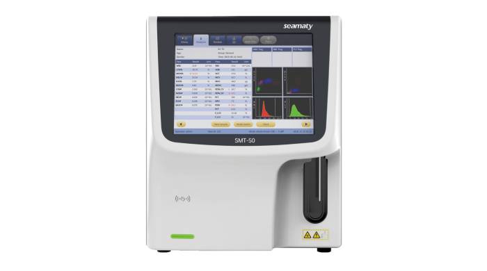
A CBC test machine is a type of medical equipment that is used to measure the levels of red blood cells, white blood cells, and platelets in a person's blood. This information is important in order to diagnose and treat various medical conditions.

3 Technologies Help Seamaty Biochemical Analyzers Be Sold Overseas

2022-09-22

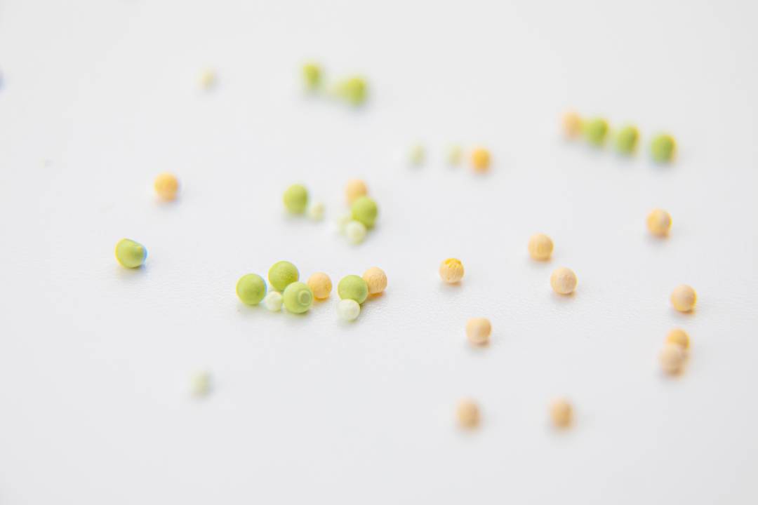
Seamaty's products are sold to Asia, Africa, Europe and America thanks to 3 core technologies: automatic calibration technology, ultrasonic welding technology and reagent lyophilization technology.

Accuracy of Blood Cell Analyzer: How the Physiological State, Analyzer and Reagents Affect

2022-07-22

Blood cell analyzer has high environmental requirements. The instrument should be far away from radiation, nuclear magnetic, radiotherapy gas pedal and other large medical equipment section to prevent the interference of electrical signals.