Whole blood is a mixture of blood cells and fluid. A complete blood count (CBC) examines the cellular fraction of the blood and gives the number of different types of red and white blood cells, platelets, and hemoglobin. Biochemical tests are performed on the fluid portion of the blood after the cells have been removed.
To obtain the fluid in the blood, the blood sample is first coagulated in a tube and then centrifuged in a centrifuge to allow the clot to sink to the bottom of the tube. The precipitated fluid remains in the upper part of the tube. The liquid that is released after clotting is called "serum" and is commonly used for biochemical assays.
Animal biochemical testing involves examining multiple chemical components within a single sample. The concentration of these chemicals can reveal information about many aspects of various organs in the body.
Veterinary clinical biochemical tests usually include the following.
Interpretation of 16 Items of Veterinary Clinical Biochemical Tests
1. Total Protein (T-Pro)
Generally speaking, total protein concentration detects two blood protein molecules, albumin and globulin.
An elevated total protein concentration is usually seen when the body is dehydrated. Globulin = total protein - albumin. The term "globulin" includes immunoglobulins. Immunoglobulins are produced by the body's immune system and protect the body from bacteria and viruses. Elevated immunoglobulin concentrations are seen in certain diseases, such as feline infectious peritonitis.
2. Albumin (Alb)
Albumin is synthesized in the liver and is the main component that makes up plasma proteins. It is broken down in peripheral tissues and has a half-life of 7-10 days. In dogs, the half-life of albumin is approximately 8.2 days. The main functions of albumin are to maintain plasma osmolarity and to act as a carrier for the transport of various hormones, ions and drugs. Elevated albumin is seen in dehydration and decreased due to decreased production, small intestine malabsorption, excessive protein loss or excessive protein breakdown.
3. Glutathione aminotransferase (GPT)
Serum ghrelin (GPT) is also known as "alanine aminotransferase" (ALT). It is an important enzyme for liver function. Elevated activity usually means that the liver cells are damaged for some reason. The liver may become cancerous, infected, congested (seen in heart failure), sclerotic, blocked (so metabolic waste and toxins in the blood cannot be removed by the bile ducts), etc. In general, any factor that adversely affects the liver or liver function will result in increased GPT activity.
4. Glutathione aminotransferase (aspartate aminotransferase)
Glutamic oxaloacetic transaminase (GOT) is also called aspartate aminotransferase (AST). It is found in many tissues, including liver, skeletal muscle, heart muscle and red blood cells. There are two isozymes, ASTs and ALTm, which are found in the cytoplasm and mitochondria, respectively. ASTs are significantly elevated during mild cellular injury. In contrast, in severe injury, ASTm is present in large amounts in the serum. In liver disease, the level of AST varies with the level of ALT. increased AST is also seen in muscle necrosis and hemolysis.
5. Alkaline phosphatase (ALP)
Serum alkaline phosphatases are enzyme molecules. These protein molecules catalyze various chemical reactions. Although reference values for alkaline phosphatase vary in various animals, elevated alkaline phosphatase in dogs is commonly seen in certain cancers and in some muscle and liver diseases.
6. Creatine phosphokinase (CK)
Elevated creatine phosphokinase activity arises from reversible damage or necrotic leakage from myocytes. Its half-life is short, and CK begins to decline within 1-2 days after cell leakage ceases. cK activity declines more rapidly than AST.
The three isozymes are present in skeletal muscle, cardiac muscle and central nervous system (CNS). A sensitive indicator of muscle injury. A peak occurs 6-12 hours after injury and returns to normal 2-3 days after injury has ceased. Persistent elevation indicates progressive or active disease.
7. Lactate dehydrogenase (LDH)
Lactate dehydrogenase is present in large amounts in all organs and tissues (including red blood cells). It is present in the cell plasma. It is released into the blood stream in response to changes in cell membrane permeability and cell necrosis. Therefore, its elevation is nonspecific. It includes 5 isoenzymes and is present in skeletal muscle, cardiac muscle, liver and blood erythrocytes. It is not organ specific and can be used to evaluate muscle damage.
8. Amylase (Amy)
Amylase is an exocrine pancreatic enzyme. It is released into the blood and peritoneal cavity after pancreatic necrosis, so large amounts of amylase can be released when the pancreas is inflamed. Other tissues containing amylase include the liver and intestinal mucosa. In addition to heparin, other anticoagulants such as citrate, oxalate and EDTA have inhibitory effects. Therefore, amylase should not be measured with decalcified plasma.
9. γ-glutamyl transferase (GGT)
GGT is present in the liver, pancreas, kidney and intestine. Elevated serum concentrations are usually caused by increased hepatic production and biliary tract disease. The highest levels are found in renal tubular epithelial cells, hepatocyte tubular surfaces and biliary epithelium. Renal tubular nephropathy and nephrotoxicity can cause elevated urinary GGT concentrations, but not serum concentrations.
10. blood glucose (Glu)
After the body ingests carbohydrates, it converts them into glycogen for storage in the liver. When the body needs energy, glycogen is converted into glucose, which enters the bloodstream and is transferred to the whole body. Therefore, blood glucose is an indicator to monitor the nutrition level of animals. However, it is more often used to detect metabolic and physiological status.
Hypoglycemia is a common disorder in young animals, especially in toy and smaller breeds. Animals suffering from hypoglycemia exhibit weakness, ataxia, and even seizures. Hypoglycemia can also occur in some adult dogs, especially with prolonged over-intense exercise, a condition commonly seen in hounds. Hypoglycemia is also seen in dogs with prolonged illness and weakness, as well as in dogs with certain cancers.
A slight increase in blood glucose can be caused by excitement or stress of the animal during blood collection. However, when the blood glucose exceeds 180 mg/dl, it is often indicative of disease. At this point, the blood glucose has exceeded the renal glucose threshold. (When blood glucose is filtered through the kidneys, the kidneys prevent the loss of glucose through the urine by reabsorption. However, once the blood glucose exceeds the renal threshold, the kidneys are unable to reabsorb all the glucose that is filtered, and glucose is then excreted in the urine). This is the common cause of diabetes. The essence of this disease is the body's inability to produce adequate amounts of insulin. Insulin is necessary for the entry of glucose into the body's cells. When insulin production is insufficient, blood sugar is retained in the blood. In diabetes, blood glucose can be as high as 900mg/dl!
10. Total bilirubin (T-Bil)
Bilirubin is a breakdown product of hemoglobin. Hemoglobin is the molecule within red blood cells that carries oxygen to the body's tissues. After death or destruction of blood cells, hemoglobin is released and quickly degraded and excreted by the liver. Therefore, when too many red blood cells are destroyed, or when the liver becomes diseased and cannot remove bilirubin from the blood, the bilirubin concentration will be higher than normal. If the liver or bile ducts become blocked and bilirubin cannot be released into the intestine, the bilirubin concentration will also increase. Doctors can combine this with direct bilirubin for a differential diagnosis.
11. Triglycerides (TG)
Triglycerides can be absorbed and synthesized in the liver to form lipoproteins along with cholesterol, phospholipids and serum proteins. Triglycerides are a major component of celiac disease and very low density lipoprotein (VLDL), the cause of lipemia, which is often seen in serum or plasma samples. After the serum/plasma sample is frozen overnight, the celiac particles separate from the serum/plasma and form a fatty layer. While VLDL remains dispersed in the serum/plasma. When an animal is fasted for 16 hours, its serum sample is determined to have pathological lipemia if it shows lipemia.
Increased triglycerides (lipemia): diabetes, hypothyroidism, acute pancreatitis, hyperadrenalism/glucocorticoid therapy, primary hyperlipoproteinemia (miniature schnauzer or beagle).
12. Total cholesterol (T-Cho)
Cholesterol is less important than human medicine in detecting disease in veterinary medicine. This is because cardiovascular sclerosis and embolism are uncommon in dogs and cats. However, changes in cholesterol concentrations are seen in other diseases as well. Elevated cholesterol concentrations are common in animals suffering from thyroid deficiency. Decreased cholesterol concentrations are seen in starved or malnourished animals.
13. Urea Nitrogen (BUN)
Dietary protein digested by the animal is a large molecule. As the protein molecule is digested and utilized by the body, its metabolic by-product urea is produced. It is useless to the organism and is eliminated via the kidneys. If the kidneys are not able to efficiently filter these substances from the blood, they will accumulate in the blood and reach very high concentrations.
A higher than normal urea nitrogen test implies that the body is not eliminating protein metabolic waste effectively. Although kidney disease is the primary cause of elevated urea nitrogen concentrations, there are other causes of elevated values. For example, a significant increase in blood urea nitrogen concentration can be seen when the body is dehydrated. This is because the body does not have enough fluid for the kidneys to work properly.
In addition, any factor that causes a decrease in blood flow to the kidneys can lead to an increase in blood urea nitrogen concentrations. For example, heart disease that causes a decrease in circulating blood flow. If there is a blockage in the urinary tract, urine cannot leave the body and accumulates in the bladder, preventing the kidneys from continuing to produce urine. This condition can also cause elevated urea nitrogen.
Blood urea nitrogen concentrations below reference values are commonly associated with liver disease. The liver is one of the main sites of protein breakdown. If catabolism does not take place, the body's nitrogenous waste concentration will fall below the normal reference value.
14. Creatinine (CRE)
Creatinine is also used to test the kidney filtration rate. Only the kidneys are capable of excreting creatinine. If it accumulates in the body above the normal reference value, it indicates decreased or impaired kidney function.
15. Calcium (Ca)
Calcium concentration in the blood is continuously constant. A significant decrease in blood calcium concentration can occur during pregnancy or lactation in dogs, a condition known as eclampsia. In addition, certain drugs and tumors can affect blood calcium concentrations. It is important to detect abnormal blood calcium promptly and quickly before it causes serious heart and muscle disease.
16. Inorganic phosphorus (P)
P is the main component of bones and teeth (where 80-85% of phosphorus is stored) and the main intracellular anion. p plays a very important role in energy metabolism and acid-base balance. It is mainly regulated by parathyroid hormones and excreted through the kidneys. p is an indicator of skeletal disease, parathyroid function and renal function.
Summary
Blood biochemical tests can provide more information than a complete blood count, which detects the cellular fraction of the blood.
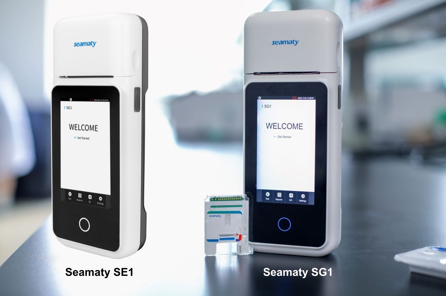
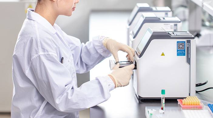
The above tests provide a direct evaluation of liver, kidney, heart, immune system and other functions. In addition to helping veterinarians with diagnosis, biochemical tests also help us determine prognosis. However, it is difficult for pet owners to afford if all tests are performed, but when performed in certain combinations, the cost of testing will become more reasonable. A dry fully automated biochemical analyzer with a combination of test strips for liver function, kidney attack, cardiovascular function, and routine tests is available. The combination of tests not only saves money, it also makes the diagnosis of the disease more comprehensive and easier.


