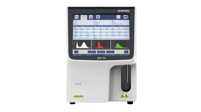
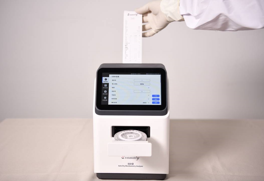
CBC machine is also called
hematology analyzer, hemocytometer, blood cell counter, etc. cbc machine is one of the most widely used instruments in hospital clinical testing. The analyzer classifies the white blood cells, red blood cells and platelets in the blood by resistance method. It can also get the data related to blood such as hemoglobin concentration and red blood cell pressure volume.
1. cbc machine's WBC histogram
The histogram of WBC in normal subjects shows two clearly separated peaks. The left peak is a small cell population (predominantly lymphocytes). The right peak is the large cell population (predominantly neutrophils). Between the two peaks are intermediate cell populations (including eosinophils, basophils, and monocytes).
The small cell population peak is high and steep. The large cell population is taller and broader. The intermediate cell population is usually a flat area with a certain width. The higher the small cell population peak, the greater the lymphocyte ratio. The higher the peak of the large cell population, the greater the neutrophil ratio. In contrast, the peak of the intermediate cell population increases when eosinophils, basophils, or monocytes are increased or when pathological cells are present. For example, in leukemia, a large number of naive cells appear in the peripheral blood, mostly in and around the intermediate cell population, resulting in higher peak tips and specific histograms for different leukemia types.
The significance of cbc machine WBC
1) To determine whether the white blood cell count is interfered with by other factors.
The WBC counting process begins with the addition of a hemolytic agent to the diluted blood to destroy the red blood cells. The retained leukocytes are counted through small wells.
The following are some of the factors that affect the white blood cell count.
① Certain anemic pathological red blood cells, neonatal red blood cells are more resistant to the hemolytic agent. This can make them non-lysing or incompletely lysed and affect the white blood cell count.
② Most nucleated red blood cells within the blood. After lysis, the nuclei of the red blood cells will be counted as white blood cells;
(3) Platelets aggregate into clumps and are mistakenly counted as white blood cells, etc.
2) Determine whether the histogram meets the screening criteria and decide whether to perform smear microscopy.
The results of "classification" of leukocytes by cbc machine (especially the resistance method, which is only a volume subgroup) are only applicable to Health Screening or without obvious abnormal results of hematological examination.
In other words, histogram abnormalities can occur when there is a significant increase or decrease in leukocytes or when there are atypical or naive cells in the peripheral blood. In this case, the results of cell "classification" are inaccurate.
Therefore, the following two conditions should be met when using a blood cell analyzer to "classify" leukocytes.
① The patient's white blood cell count, platelet count, and hemoglobin content are within the normal range;
The histogram of white blood cell volume distribution is completely normal before it can be used as a clinical result of leukocyte classification, otherwise it must be smear microscopy.
2. Meaning of "R" in cbc machine
R1 (suggesting abnormalities in the area to the left of the lymphocyte peak on the leukocyte histogram): aggregated platelets, giant platelets, nucleated red blood cells, unresolved red blood cells, protein or lipid particles may be present.
R2 (indicates abnormalities in the area between the lymphocyte peak and the monocyte area on the leukocyte histogram): possible presence of heterogeneous lymphocytes, plasma cells, atypical lymphocytes, primitive cells, eosinophilia, and basophilia
R3 (suggests abnormalities in the area between the monocyte zone and the neutrophil peak on the leukocyte histogram): possible presence of immature neutrophils, abnormal cell subsets, eosinophilia.
R4 (indicates an abnormality in the area to the right of the neutrophil peak on the leukocyte histogram): indicates an increase in absolute neutrophil values.
RM (multiple site alarm): indicates the presence of two or more "R" alarms at the same time.
3. RBC histogram of cbc machine
The RBC histogram is a graph reflecting the distribution of particles within the size of the RBC volume or any range equivalent to the RBC volume size. A normal RBC histogram is a skewed distribution graph. The peak indicates the average of a certain number of RBC volumes, which is basically the same as the average RBC volume. The peak value is generally between 82-94fl in normal individuals.
The peak of the histogram becomes lower and the base widens, indicating that the RBC volume varies in size, which is consistent with the increase in the distribution of red blood cells.
A leftward shift of the histogram suggests a smaller RBC volume, such as a significant leftward shift of the RBC histogram in patients with iron deficiency anemia and a significant widening of the base.
A rightward shift of the histogram suggests a larger RBC volume. This is common in megaloblastic anemia and neonates.
The RBC histogram sometimes shows a double peak, which is evidence of effective anemia treatment, mostly seen in patients with iron deficiency anemia and megaloblastic anemia after treatment.
Significance of the RBC histogram of the cbc machine
The RBC histogram combined with the parameters is valuable for the differential diagnosis of anemia. The type of anemia can be determined by looking at the position of the graph peaks, the width of the bottom of the peaks, the shape of the top of the peaks and the presence or absence of double peaks.
4. PLT histogram of cbc machine
PLT histogram is a distribution chart reflecting the frequency of PLT volume distribution. A normal PLT histogram has a left skewed distribution with peaks generally between 6-10fl.
A left shift in the histogram indicates a small PLT volume, while a right shift in the histogram indicates a large PLT volume. An elevated tail of the histogram or another peak at 20-30fl suggests that there may be more small red blood cells, red blood cell debris, giant platelets, platelet agglutination and fibrin in the blood. The PLT histogram in patients with iron deficiency anemia has a significantly elevated tail and a decreased PLT and its histogram during treatment is mostly wavy.
Significance of PLT histogram
Since platelet measurements are in the same channel as red blood cell measurements. It is only because there is a significant difference between platelet volume and red blood cell volume that the instrument sets a specific threshold. The instrument designates those above the threshold as red blood cells and vice versa as platelets.
However, this is not actually the case. A very small percentage of the red blood cell population can fall within the threshold for platelets. And large platelets can be mistaken for red blood cells. In order to compensate for the loss of large platelets, the instrument calculates the "lost" area based on the characteristics of the platelet pattern, such as wave crest and half-wave width, as compensation for the platelet count.
However, this compensation is based on the condition that the graph is basically normal. The results of the platelet count and MPV (which are also derived from the histogram) tests are affected by various factors that make the graph significantly abnormal. Therefore, analysis of platelet histograms is an important step in the post-analytical quality control of platelet tests.
Platelet histograms are of little value to physicians as a basis for diagnosis and treatment. The platelet histogram can reflect the interference of small red blood cells, large platelets, red blood cell debris and platelet aggregation with the platelet count and MPV value of the specimen being tested.
The significance of the blood test is to detect early signs of many systemic diseases. The analyzer can diagnose whether there is anemia, whether there is a blood disorder, reflect the hematopoietic function of the bone marrow, etc. Therefore, in laboratory medicine, blood cell analyzers assume a very important


