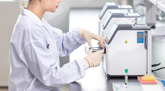
There are several chemical analysis techniques in biochemistry analyzers, including colorimetric reaction, ISE (ion selective electrode), immunoturbidimetry, etc. The way and container of colorimetric reaction is the main difference between dry and wet biochemistry analyzer.
The main difference between dry and wet biochemistry analyzers is the way and the vessel in which the colorimetric reaction occurs. Accordingly, the optical path systems of the two types of biochemical analyzers are also different.
Wet biochemistry analyzer
Normal wet biochemistry analyzers generally have color development reactions in a colorimetric cup or flow cell. Biochemistry analyzers that usually use a colorimetric cup are also equipped with a stirrer. This ensures an adequate and rapid reaction.
Therefore, a normal wet cell biochemistry analyzer must be equipped with a piping system for sample aspiration, sample dilution, reagent distribution and other functions.
Piping systems for flow cell biochemistry analyzers also need to perform functions such as pipeline cleaning and drainage of waste liquids. In addition, ordinary wet biochemical analyzers usually use photoelectric colorimetric method for analysis, and its absorbance and the concentration of the measured solution satisfy Lambert-Bier's law.
Dry biochemical analyzer
Dry biochemical analyzers, on the other hand, completely abandon the traditional pipeline system and use dry reagent strips as solid phase reagents. Dry reagent strips are generally composed of multiple layers of film.
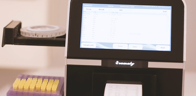
For example, some biochemical analyzers have a multilayer film composition of dry reagents as follows.
-
The first layer is the distribution diffusion layer: it can make the specimen evenly distributed and can filter large molecules. Minimizes hemolysis, hyperlipidemia and bilirubin interference. Also provides a background for reflectometry.
-
The second layer is the reagent layer: contains dry reagents. Controls the reaction sequence.
-
The third layer is the indicator layer: contains the dyes to produce the color development complex.
The multilayer film technique not only masks the colored material to be measured and provides a background, selectively retains or removes interfering material. It also carries out a variety of substances in layers equivalent to the reaction principle of the wet chemical method.
The reaction products in one layer can in turn enter another layer for other reactions, thus directing the reaction sequence. Each layer can give a specific environment to control the reaction sequence and reaction time. The concentration of the components to be measured in the sample can be calculated by the Kubelka-Munk theory.
These are the structural differences between dry and wet biochemical analyzers.